X-ray method of examination of the patient. Basic methods of X-ray examination. Indications for bone x-ray
Send your good work in the knowledge base is simple. Use the form below
Students, graduate students, young scientists who use the knowledge base in their studies and work will be very grateful to you.
Posted on http://www.allbest.ru/
Tema. X-ray research methods
x-ray microscopy beam spectroscopy
X-ray radiation discovered (1895) by a German physicist, Nobel Laureate(1901) W. Röntgen, occupies the spectral region between gamma and UV radiation within wavelengths of 10-3-102 nm. Radiation with< 0,2 нм условно называют жестким, а с >0.2 nm - soft. The totality of X-ray research methods includes X-ray microscopy, spectroscopy, and X-ray structural and phase analyses.
X-ray spectroscopy
X-ray spectroscopy (X-ray spectral analysis) studies x-ray spectra of emission (emission spectroscopy) and absorption (absorption spectroscopy).
X-ray spectra are a consequence of electron transitions in the inner shells of atoms. To obtain X-ray spectra, the sample is bombarded with electrons in an X-ray tube (electrovacuum device for obtaining x-rays) or excite the fluorescence of the test substance by irradiating it with X-rays. Flow primary x-ray radiation is directed to the sample, and the secondary X-ray radiation reflected from it falls on the analyzer crystal. X-ray diffraction is carried out on its atomic structure - the decomposition of secondary radiation into a spectrum along wavelengths. The reflected flow is sent for registration (X-ray film, ionization chamber, counter, etc.).
X-ray absorption spectra carry information about the transition of electrons from the inner shell of an atom to excited shells. The spectrum has a sharp boundary (absorption threshold) in the region of low radiation frequencies. The part of the spectrum before it corresponds to electron transitions to bound states. Beyond the absorption threshold, the interaction of electrons removed from an atom with neighboring atoms leads to the appearance of absorption minima and maxima in the spectrum. The distances between them correlate with the interatomic distances in the sample material.
X-ray emission spectra (emission spectra) carry information about the transition of electrons from valence shells to vacancies in the inner shells, i.e. reflect the structure of the valence shells of the atom. Particularly valuable information is obtained by analyzing the dependence of the intensity of lines in the emission spectra of a single crystal on the angle of rotation of the sample. In this case, the line intensities are proportional to the population of the levels from which the electron transition takes place.
According to the signs of the excitation mechanism of the primary radiation incident on the sample, three methods of X-ray spectroscopy are distinguished: X-ray spectral microanalysis, X-ray fluorescence and X-ray radiometric analysis.
X-ray spectral microanalysis is based on the excitation by an electron probe (beam of focused electrons) of characteristic X-ray radiation in a sample. An electron probe (diameter ~ 1 μm) is formed using X-ray microanalyzers based on electron microscopes (transmission or scanning). The instrument is under high vacuum. Atomic numbers are identified from the spectrum of the characteristic X-ray radiation excited by the probe on a microsection of the sample. chemical elements, and according to the intensity of the lines - their concentration in the microsection. The absolute and relative limits of detection of elements in the sample are 10-12-10-6 g and 10-1-10-3%, respectively.
X-ray fluorescence analysis (XRF) is based on the use of secondary X-rays to eliminate radiation damage to the sample and increase the reproducibility of results. The device consists of an x-ray tube, an analyzer crystal that decomposes the secondary radiation into a spectrum, and a detector - a counter of ionizing radiation.
Qualitative XRF is based on the analysis of the dependence of the frequency of the characteristic X-ray radiation emitted by a chemical element on the atomic number of the element. XRF is designed to study chemical bonds, distribution of valence electrons, determination of the charge of ions. It is used in the analysis of materials in metallurgy, geology, in the processing of ceramics, etc.
X-ray radiometric analysis (XRA) involves the measurement of X-ray radiation, which occurs when the radiation of a radioisotope source interacts with electrons located on the inner shells of the atoms of the analyzed substance. With the fluorescent version of the method, the flux of X-ray fluorescence quanta is measured, the energy of which characterizes the chemical element, and the intensity characterizes its content. The absorption variant provides for recording the attenuation of two X-ray fluxes with similar energies by the sample. The ratio of the intensities of the flows that have passed through the sample characterizes the content of the element being determined.
The PPA method allows elemental analysis of mixtures and surface layers solids. The limit of detection is 10-4-10-10%, the duration of the determination is within 10 minutes. PPA analyzers have been used to study the elemental composition of rocks on the Moon and Venus.
X-ray spectroscopy methods include a method that is at the junction of X-ray and electron spectroscopy.
X-ray electron spectroscopy (XES), or electron spectroscopy for chemical analysis(ESCA), allows you to study the electronic structure of chemical compounds, the composition and structure of the surface layer of solids using the photoelectric effect caused by X-rays. An analysis of the kinetic energy of electrons emitted from a sample provides information about the elemental composition of the sample, the distribution of chemical elements on its surface, the nature of chemical bonds, and other interactions of atoms in the sample.
In electron spectrometers, the sample is usually exposed to X-ray tube radiation. The electrons e, knocked out by the X-ray quantum, enter the electronic energy analyzer, which separates them by energy. Monochromatic electron beams are directed to a detector that measures the intensity of the beams. As a result, an x-ray electron spectrum is obtained - the distribution of x-ray photoelectrons by kinetic energies. The maxima on it (spectral lines) correspond to certain atoms. X-ray electron spectroscopy is one of the main methods for determining the composition of the surface layers of bodies; it is widely used in the study of adsorption, catalysis, and corrosion. This is one of the main methods for determining the thickness and continuity of single-crystal thin films.
X-ray structural analysis
X-ray structural analysis (XRD) is a set of methods for studying the atomic structure of a substance, mainly crystals, using X-ray diffraction. It is based on the interaction of X-ray radiation with the electrons of the substance under study, resulting in diffraction. Its parameters depend on the wavelength of the radiation used and atomic structure object. According to the diffraction pattern, the distribution of the electron density of the substance is established, and according to it, the type of atoms and their arrangement in the crystal lattice. To study the atomic structure, radiation with a wavelength of ~ 0.1 nm is used, i.e. about the size of an atom.
Since the 1950s, computers have been used in the processing of X-ray diffraction patterns.
For X-ray structural analysis, X-ray cameras, diffractometers and goniometers are used.
An X-ray camera is a device for studying and controlling the atomic structure of substances, which uses X-ray tube radiation and creates conditions for X-ray diffraction on a sample, and the diffraction pattern is recorded on photographic film.
X-ray diffractometer - a device for X-ray structural analysis, which is equipped with photoelectric radiation detectors. It measures the intensity and direction of diffractive X-ray beams.
X-ray goniometer - a device for X-ray structural analysis, which simultaneously registers the direction of diffraction rays and the position of the sample.
Scattered X-rays are recorded on photographic film or measured using nuclear radiation detectors, which are based on the phenomena that occur when charged particles pass through matter. To register the formed particles, ionization chambers, counters, semiconductor detectors are used, and for visual observation and photographing traces (tracks) of particles, track detectors (nuclear photographic emulsions, bubble and spark chambers, etc.) are used. A diffraction pattern can be created in several ways. Their choice is determined by the physical state and properties of the sample, as well as the amount of information that needs to be obtained about it.
The Laue method is the simplest method for obtaining x-ray patterns from single crystals: the sample is fixed motionless, x-ray radiation has a continuous spectrum. An x-ray pattern containing a diffraction image of a single crystal is called a Laue pattern. The location of diffraction spots on it depends on the symmetry of the crystal and its orientation relative to the primary beam. By the manifestation of asterism - blurring in certain directions of diffraction spots on Laue patterns - stresses in the sample and some crystal defects are revealed.
Sample rocking and rotation methods are used to determine the unit cell parameters in a crystal. The diffraction pattern created by monochromatic radiation is recorded on an x-ray film located in a cylindrical cassette, the axis of which coincides with the axis of oscillation of the sample. The diffraction spots on the unfolded film are located on the family parallel lines. Knowing the distance between them, the cassette diameter and the radiation wavelength, the parameters of the crystal cell are calculated.
X-ray goniometric methods are designed to measure the parameters of diffraction reflections from a crystal for all possible orientations. The intensity of reflections is determined: photographically, measuring the degree of blackness of each spot on the radiograph with a microphotometer; directly with the help of X-ray counters.
A series of radiographs is obtained in X-ray goniometers. Each of them recorded diffraction reflections, the crystallographic indices of which have certain limitations. When studying a structure consisting of ~ 50-100 atoms, it is necessary to measure the intensity of about 100-1000 diffraction reflections. This time-consuming and painstaking work is carried out with the help of computer-controlled multichannel diffractometers.
The Debye-Scherrer method for studying polycrystals consists in recording scattered radiation on a photographic film (Debyegram) in a cylindrical X-ray chamber. The Debyegram of a polycrystal consists of several concentric rings and makes it possible to identify chemical compounds, determine the phase composition of samples, grain sizes and texturing, and control stresses in a sample.
The small-angle scattering method makes it possible to detect spatial inhomogeneities in condensed bodies, the dimensions of which (from 0.5 to 103 nm) exceed the interatomic distances. The small-angle scattering method is used to study nanocomposites, metal alloys, and complex biological objects. It has proven to be effective for the industrial control of catalysts.
X-ray topography, which is sometimes referred to as methods of X-ray structural analysis, makes it possible to study defects in the structure of almost perfect crystals by studying the diffraction of X-rays on them. Carrying out the diffraction of X-rays on the crystals "for transmission" and "for reflection" in special X-ray cameras, diffraction images of the crystal are recorded - a topogram. By deciphering it, they obtain information about defects in the crystal. The linear resolution of X-ray topography methods is from 20 to 1 microns, the angular resolution is from 1" to 0.01""
Based on the results of their X-ray structural analysis, it is possible to determine the atomic structure of crystals.
An analysis of the diffraction of x-rays makes it possible, in addition, to determine the quantitative characteristics of the thermal vibrations of atoms in a crystal and the spatial distribution of electrons in it. Laue and sample rocking methods measure the parameters of the crystal lattice. When studying a single crystal, the shape and dimensions of the unit cell of the crystal are determined from the diffraction angles. According to the regular absence of some reflections, one judges the space group of symmetry. The intensity of the reflections is used to calculate the absolute values of the structural amplitudes, which are used to judge the thermal vibrations of atoms. Calculations are carried out using a computer.
To solve many problems of physics, chemistry, molecular biology and others. The joint use of methods of X-ray diffraction analysis and resonance methods (EPR, NMR, etc.) is effective.
X-ray phase analysis
X-ray phase analysis is a method of qualitative and quantification phase composition of polycrystalline materials, based on the study of X-ray diffraction.
Qualitative X-ray phase analysis is aimed at determining the distance between parallel crystallographic planes. By its value, the chemical nature of the investigated crystalline phase is identified by comparing the obtained value with the known values of this distance for individual phases. The phase is considered to be established if there are three of its most intense peaks on the diffraction pattern and the approximate correspondence of the ratio of their intensities to the reference data.
Quantitative X-ray phase analysis of a mixture of two phases is based on the dependence of the ratio of the intensities of the diffraction peaks of these phases on the ratio of their concentrations. The error in the quantitative determination of the phase by this method is approximately 2%.
Hosted on Allbest.ru
...Similar Documents
Instrumental research methods in medicine with the use of apparatus, devices and instruments. Use of x-rays in diagnostics. X-ray examination of the stomach and duodenum. Methods of preparation for the study.
presentation, added 04/14/2015
The history of the discovery of X-rays by the German physicist Wilhelm Roentgen. The process of obtaining x-ray radiation, its application in medical research. Modern types of X-ray diagnostics. Computed X-ray tomography.
presentation, added 04/22/2013
Biography and scientific activity VK. Roentgen, the history of his discovery of X-rays. Characterization and comparison of two main methods in medical X-ray diagnostics: fluoroscopy and radiography. Examination of the organs of the gastrointestinal tract and lungs.
abstract, added 03/10/2013
Characteristic laboratory diagnostics viral infections using electron microscopy. Preparation of sections of affected tissue for examination. Description of the method of immunoelectron microscopy. Immunological research methods, description of the course of analysis.
term paper, added 08/30/2009
Conducting a general sputum analysis - a study for the initial assessment of the condition of the bronchi and lungs. Collection and analysis of sputum. The main factors influencing the result of the study. Microscopy, bacterioscopy and sputum culture. Study of physical properties.
abstract, added 11/05/2010
Introduction to the history of the discovery of X-rays. Development of this diagnostics in Germany, Austria, Russia. The device and principle of operation of the x-ray tube, the properties of the rays. The device of the X-ray apparatus, the corresponding department (office).
presentation, added 02/10/2015
Approximate and quantitative method for the study of urine sediment. Calculation of the daily number of shaped elements. Unaltered and altered erythrocytes. Hyaline and granular casts. Cells of stratified squamous epithelium. Calcium oxalate crystal.
presentation, added 04/14/2014
The discovery of X-rays by Wilhelm Roentgen, the history and significance of this process in history. The device of an x-ray tube and the relationship of its main elements, principles of operation. Properties of X-ray radiation, its biological effect, role in medicine.
presentation, added 11/21/2013
Enalapril: main properties and mechanism of obtaining. Infrared spectroscopy as a method for identifying enalapril. Methods for testing the purity of a given medicinal substance. Pharmacodynamics, pharmacokinetics, use, and side effects of enalapril.
abstract, added 11/13/2012
Cytogenetic research methods. Indications for the diagnosis of hereditary pathology. Method of genomic hybridization. Cytogenetic localization of DNA sequences. The main indications in newborns and children. Magnetic resonance spectroscopy.
Electron-optical amplification (EOA). The operation of an electron-optical converter (IOC) is based on the principle of converting an X-ray image into an electronic image with its subsequent transformation into an amplified light image. The brightness of the screen glow is enhanced up to 7 thousand times. The use of an EOS makes it possible to distinguish details with a size of 0.5 mm, i.e. 5 times smaller than with conventional fluoroscopic examination. When using this method, X-ray cinematography can be used, i.e. recording an image on film or videotape.
Radiography is photography using x-rays. When taking X-rays, the object to be photographed must be in close contact with the cassette loaded with film. X-ray radiation coming out of the tube is directed perpendicularly to the center of the film through the middle of the object (the distance between the focus and the patient's skin under normal operating conditions is 60-100 cm). Indispensable equipment for radiography are cassettes with intensifying screens, screening grids and a special x-ray film. The cassettes are made of opaque material and correspond in size to the standard sizes of produced X-ray film (13 × 18 cm, 18 × 24 cm, 24 × 30 cm, 30 × 40 cm, etc.).
Intensifying screens are designed to increase the light effect of x-rays on photographic film. They represent cardboard, which is impregnated with a special phosphor (calcium tungsten acid), which has a fluorescent property under the influence of X-rays. Currently, screens with phosphors activated by rare earth elements are widely used: lanthanum oxide bromide and gadolinium oxide sulfite. The very good efficiency of the rare earth phosphor contributes to the high light sensitivity of the screens and ensures high image quality. There are also special screens - Gradual, which can even out the existing differences in the thickness and (or) density of the subject. The use of intensifying screens significantly reduces the exposure time for radiography.
Special movable gratings are used to filter out the soft rays of the primary flux that can reach the film, as well as the secondary radiation. Processing of the filmed films is carried out in a photo laboratory. The processing process is reduced to development, rinsing in water, fixing and thorough washing of the film in flowing water, followed by drying. Drying of films is carried out in drying cabinets, which takes at least 15 minutes. or occurs naturally, with the picture being ready the next day. When using processing machines, images are obtained immediately after the study. Advantage of radiography: eliminates the disadvantages of fluoroscopy. Disadvantage: the study is static, there is no possibility of assessing the movement of objects during the study.
Electroroentgenography. Method for obtaining x-ray images on semiconductor wafers. The principle of the method: when rays hit a highly sensitive selenium plate, the electric potential changes in it. The selenium plate is sprinkled with graphite powder. Negatively charged powder particles are attracted to those areas of the selenium layer in which positive charges have been preserved, and are not retained in those areas that have lost their charge under the action of X-rays. Electroradiography allows you to transfer the image from the plate to paper in 2-3 minutes. More than 1000 shots can be taken on one plate. The advantage of electroradiography:
1. Speed.
2. Profitability.
Disadvantage: insufficiently high resolution in the study of internal organs, a higher dose of radiation than with radiography. The method is used mainly in the study of bones and joints in trauma centers. Recently, the use of this method has been increasingly limited.
Computed X-ray tomography (CT) (Appendix 2). The creation of X-ray computed tomography was major event in radiodiagnosis. This is evidenced by the award Nobel Prize in 1979 to renowned scientists Cormac (USA) and Hounsfield (England) for the development and clinical testing of CT.
CT allows you to study the position, shape, size and structure of various organs, as well as their relationship with other organs and tissues. Various models of mathematical reconstruction of X-ray images of objects served as the basis for the development and creation of CT. Advances achieved with the help of CT in the diagnosis of various diseases served as a stimulus for the rapid technical improvement of devices and a significant increase in their models. If the first generation of CT had one detector, and the time for scanning was 5-10 minutes, then on tomograms of the third - fourth generations, with 512 to 1100 detectors and high-capacity computers, the time to obtain one slice decreased to milliseconds, which practically allows you to explore all organs and tissues, including the heart and blood vessels. Currently, spiral CT is used, which makes it possible to carry out a longitudinal reconstruction of the image, to study rapidly occurring processes (contractile function of the heart).
CT is based on the principle of creating an x-ray image of organs and tissues using a computer. CT is based on the registration of X-ray radiation by sensitive dosimetric detectors. The principle of the method lies in the fact that after the rays pass through the patient's body, they do not fall on the screen, but on the detectors, in which electrical impulses arise, transmitted after amplification to the computer, where, according to a special algorithm, they are reconstructed and create an image of the object that is fed from the computer on a TV monitor. The image of organs and tissues on CT, unlike traditional x-rays, is obtained in the form of transverse sections (axial scans). With helical CT, a three-dimensional image reconstruction (3D mode) with high spatial resolution is possible. Modern installations make it possible to obtain sections with a thickness of 2 to 8 mm. The X-ray tube and radiation receiver move around the patient's body. CT has a number of advantages over conventional X-ray examination:
1. First of all, high sensitivity, which makes it possible to differentiate individual organs and tissues from each other in terms of density up to 0.5%; on conventional radiographs, this figure is 10-20%.
2. CT allows you to get an image of organs and pathological foci only in the plane of the examined section, which gives a clear image without layering the formations lying above and below.
3. CT makes it possible to obtain accurate quantitative information about the size and density of individual organs, tissues and pathological formations.
4. CT makes it possible to judge not only the state of the organ under study, but also the relationship of the pathological process with surrounding organs and tissues, for example, tumor invasion into neighboring organs, the presence of other pathological changes.
5. CT allows you to get topograms, i.e. a longitudinal image of the area under study, like an x-ray, by moving the patient along a fixed tube. Topograms are used to establish the extent of the pathological focus and determine the number of sections.
6. CT is indispensable for radiotherapy planning (radiation mapping and dose calculation).
CT data can be used for diagnostic puncture, which can be successfully used not only to detect pathological changes, but also to assess the effectiveness of treatment and, in particular, antitumor therapy, as well as to determine relapses and associated complications.
Diagnosis by CT is based on direct radiographic features, i.e. determining the exact localization, shape, size of individual organs and the pathological focus and, most importantly, on indicators of density or absorption. The absorbance index is based on the degree to which an X-ray beam is absorbed or attenuated as it passes through the human body. Each tissue, depending on the density of the atomic mass, absorbs radiation differently, therefore, at present, the absorption coefficient (HU) on the Hounsfield scale has been developed for each tissue and organ. According to this scale, HU of water is taken as 0; bones with the highest density - for +1000, air with the lowest density - for -1000.
The minimum size of a tumor or other pathological focus, determined by CT, ranges from 0.5 to 1 cm, provided that the HU of the affected tissue differs from that of healthy tissue by 10-15 units.
In both CT and X-ray studies, it becomes necessary to use the “image enhancement” technique to increase the resolution. Contrast in CT is performed with water-soluble radiopaque agents.
The “enhancement” technique is carried out by perfusion or infusion administration of a contrast agent.
Such methods of X-ray examination are called special. The organs and tissues of the human body become visible if they absorb x-rays to varying degrees. Under physiological conditions, such differentiation is possible only in the presence of natural contrast, which is determined by the difference in density ( chemical composition of these organs), size, position. The bone structure is well detected against the background of soft tissues, the heart and large vessels against the background of airy lung tissue, however, the chambers of the heart under conditions of natural contrast cannot be distinguished separately, as well as the organs of the abdominal cavity, for example. The need to study organs and systems with the same density by X-rays led to the creation of a technique for artificial contrasting. The essence of this technique is the introduction of artificial contrast agents into the organ under study, i.e. substances having a density different from the density of the organ and its environment.
X-ray contrast media (RCS) are usually divided into substances with high atomic weight (X-ray positive contrast agents) and low (X-ray negative contrast agents). The contrast agents must be harmless.
Contrast agents that absorb intensely x-rays (positive radiopaque agents) are:
1. Suspensions of salts of heavy metals - barium sulfate, used to study the gastrointestinal tract (it is not absorbed and excreted through natural routes).
2. Aqueous solutions organic compounds of iodine - urographin, verografin, bilignost, angiographin, etc., which are introduced into the vascular bed, enter all organs with the blood flow and give, in addition to contrasting the vascular bed, contrasting other systems - urinary, gallbladder, etc.
3. Oily solutions of organic compounds of iodine - iodolipol, etc., which are injected into fistulas and lymphatic vessels.
Non-ionic water-soluble iodine-containing radiopaque agents: ultravist, omnipak, imagopak, vizipak are characterized by the absence of ionic groups in the chemical structure, low osmolarity, which significantly reduces the possibility of pathophysiological reactions, and thereby causes a low number of side effects. Non-ionic iodine-containing radiopaque agents cause a lower number of side effects than ionic high-osmolar contrast media.
X-ray negative or negative contrast agents - air, gases "do not absorb" x-rays and therefore shade well the organs and tissues under study, which have a high density.
Artificial contrasting according to the method of administration of contrast agents is divided into:
1. The introduction of contrast agents into the cavity of the organs under study (the largest group). This includes studies of the gastrointestinal tract, bronchography, fistula studies, all types of angiography.
2. The introduction of contrast agents around the organs under study - retropneumoperitoneum, pneumothorax, pneumomediastinography.
3. The introduction of contrast agents into the cavity and around the organs under study. This includes parietography. Parietography in diseases of the gastrointestinal tract consists in obtaining images of the wall of the investigated hollow organ after the introduction of gas, first around the organ, and then into the cavity of this organ. Usually, parietography of the esophagus, stomach and colon is performed.
4. A method based on the specific ability of some organs to concentrate individual contrast agents and at the same time shade it against the background of surrounding tissues. These include excretory urography, cholecystography.
Fluorography is a method of mass in-line X-ray examination, which consists in photographing an X-ray image from a translucent screen onto a film with a camera.
Tomography (conventional) - to eliminate the summation nature of the x-ray image. Principle: during the shooting process, the X-ray tube and film cassette move synchronously relative to the patient. As a result, a clearer image of only those details that lie in the object at a given depth is obtained on the film, while the image of the details located above or below becomes blurred, “smeared”.
Polygraphy is the obtaining of several images of the organ under study and its part on one radiograph. Several shots are taken (mostly 3) on one film after a certain time.
X-ray kymography is a method of objective registration of the contractility of the muscle tissue of functioning organs by changing the contour of the image. The picture is taken through a moving slit-like lead grating. In this case, the oscillatory movements of the organ are recorded on the film in the form of teeth that have a characteristic shape for each organ.
Digital radiography - includes the detection of a beam pattern, image processing and recording, image presentation and viewing, information storage. With this technology, the detector converts the X-ray radiation after it passes through the object under study into an electrical signal, which in the analog-to-digital converter "turns" into numerical values. Computer processing of the resulting digital image serves to create an image that is optimally suitable for analyzing the examination result.
X-ray diagnostics - medical and diagnostic procedures. This refers to combined X-ray endoscopic procedures with medical intervention. For example: with obstructive jaundice with drainage of the biliary tract and the introduction of drugs directly into the gallbladder. X-ray diapeutics (interventional radiology) includes X-ray endovascular interventions: X-ray endovascular occlusion and X-ray endovascular dilatation.
Ultimately, the subject of study in radiology is the shadow image. The features of the shadow x-ray image are:
1. An image consisting of many dark and light areas - corresponding to areas of unequal attenuation of X-rays in different parts object.
2. The dimensions of the X-ray image are always enlarged (except for CT) compared to the object under study, and the larger the further the object is from the film, and the smaller the focal length (distance of the film from the focus of the X-ray tube).
3. When the object and film are not in parallel planes, the image is distorted.
4. Summation image (except tomography). Therefore, x-rays must be made in at least two mutually perpendicular projections.
5. Negative image on X-ray and CT.
Each tissue and pathological formations detected by radiological examination are characterized by strictly defined features, namely: number, position, shape, size, intensity, structure, nature of the contours, the presence or absence of mobility, dynamics over time.
Application in medicine
Radiography is used for diagnosis: X-ray examination (hereinafter referred to as RI) of organs allows you to clarify the shape of these organs, their position, tone, peristalsis, and the state of the relief of the mucous membrane.
· RI of the stomach and duodenum (duodenography) is important for the recognition of gastritis, ulcerative lesions and tumors.
X-ray examination of the duodenum is an important auxiliary method for diagnosing pathological changes in the common bile duct and the major duodenal papilla (MDP). More clearly, the pathological process can be identified when conducting an x-ray examination of the duodenum in conditions of its relaxation, called relaxation, or hypotonic duodenography. This method of studying the duodenum is highly appreciated by domestic and foreign researchers.
Relaxation duodenography allows diagnosing the tumor process of duodenal duodenal dysplasia, as well as the head of the pancreas, and confirming the mechanical cause of developed jaundice. In patients in whom surgery on the biliary tract ended with the formation of bilioduodenal anastomoses, it gives an idea of the function of the formed anastomosis and reveals pathological processes in the hepatobiliary duct, which cause relapses of suffering.
RI of the gallbladder (cholecystography) and biliary tract (cholegraphy) is carried out to assess the contours, size, lumen of the intra- and extrahepatic bile ducts, the presence or absence of calculi, and clarify the concentration and contractile functions of the gallbladder.
Cholecystography is a method of X-ray examination of the gallbladder using a contrast agent. Before cholecystography, a survey x-ray of the right half of the abdominal cavity is made. 12-15 hours before cholecystography, the patient takes bilitrast or another contrast agent, drinking it with sweet tea. The night before and 2 hours before the examination, the patient is cleansed with an enema. After transillumination, several pictures of the gallbladder are taken in different projections with the vertical and horizontal positions of the subject. Then the patient eats a special breakfast (egg yolks, butter) and he takes a few more shots with an interval of 15-20 minutes.
Cholecystography allows you to determine the position, shape, size, displacement of the gallbladder, its ability to concentrate bile and contract after eating fatty foods. Cholecystography can be done on an inpatient or outpatient basis to recognize functional or organic lesions, and in particular gallbladder stones, which are seen on cholecystograms as filling defects.
· RI of the colon (irrigoscopy) is used to recognize tumors, polyps, diverticula and intestinal obstruction.
Irrigoscopy - X-ray examination of the colon with retrograde filling of its radiopaque suspension. Irrigoscopy is used to clarify the diagnosis of diseases of the colon (malformations, tumors, chronic colitis, diverticulosis, fistulas, cicatricial narrowing, etc.).
Irrigoscopy makes it possible to obtain information about the morphological changes in the colon, which is more valuable in terms of the diagnosis of nosological forms. Irrigoscopy is often decisive method diagnosis of tumors, diverticula of the colon. The double contrast technique increases the diagnostic possibilities of irrigoscopy. In relation to diseases such as colitis, tuberculosis, only indirect signs can be obtained.
X-ray of the chest organs - a classic projectional chest x-ray examination used to diagnose pathological changes in the chest, chest cavity organs and nearby anatomical structures. A chest x-ray is one of the most common x-ray studies.
As with other x-ray examinations, one of the types is used to obtain a chest x-ray. ionizing radiation- X-ray radiation.
Chest X-ray helps to identify pathological changes in soft tissues, chest bones and anatomical structures located in the chest cavity (lungs, pleura, mediastinum). Pneumonia and congestive heart failure are most commonly diagnosed on x-rays. Along with diagnostic purposes, chest radiography is used as a screening method to assess the condition of the lung tissue, in particular in persons with occupational hazards (for example, miners).
For some diseases of the chest, radiography is good as a screening method, but has insufficient diagnostic value; in these cases, additional research(computed tomography, bronchoscopy, etc.).
It should be borne in mind that in some cases, chest x-ray may not be informative (that is, show a false negative result). Such situations may be due to the projection layering of the shadow of the pathological focus on the shadow of a normal anatomical structure (for example, diaphragm, mediastinum), low intensity of the focus (for example, initial inflammatory manifestations), inadequate projection of the study (especially in the case of pathology of the mediastinum or fractures of the ribs, sternum) .
Spine - degenerative-dystrophic (osteochondrosis, spondylosis, curvature), infectious and inflammatory (various types of spondylitis), tumor diseases.
Various parts of the peripheral skeleton - for various traumatic (fractures, dislocations), infectious and tumor changes.
Abdominal cavity - perforation of organs, kidney function (excretory urography) and other changes.
Excretory_urography_an X-ray method for examining the urinary tract, based on the ability of the kidney to secrete (excrete) certain radiopaque substances introduced into the body, resulting in an image of the kidneys and urinary tract on radiographs. As a radiopaque substance, iodine-containing concentrated (60-80%) solutions of sergosin, urografin, urotrast, etc. are used. The drug is administered intravenously in a stream slowly (within 2-3 minutes). The amount of contrast is calculated per weight.
A series of radiographs taken: the first at the 5-7th, the second at the 12-15th, the third at the 20-25th minute, in case of delay in the excretion of the contrast agent, delayed pictures are taken at the 45th and 60th minutes. The study allows you to get an almost complete picture of the secretion of a contrast agent by the kidneys and its progress through the urinary tract. The number of images is determined by the type of pathology.
When analyzing excretory urograms, the position, shape, size, contours of the kidneys, the functional state of the kidneys, the shape and contours of the ureters and bladder are evaluated.
· Metrosalpingography(MSH) is one of the most commonly used methods of gynecological examination for infertility, which allows to detect obstruction of the fallopian tubes and peritubal adhesions. The most modern, least traumatic and most informative method for assessing tubal patency today is selective metrosalpingography(the introduction of contrast is carried out from the side of the uterine cavity aiming at the mouth of the fallopian tubes). Selective MSG suggests the possibility of recanalization(restoration of patency) of the fallopian tubes in violation of the patency of the interstitial (initial) sections of the tubes. The selective MSH procedure is not accompanied by pain and does not require the use of anesthesia. As a rule, it is enough to take antispasmodics and standard painkillers the day before the procedure.
· Orthopantomography
X-ray examination in dentistry, ENT, maxillofacial surgery, cosmetology, etc., which allows you to get a detailed image of all teeth with jaws, adjacent parts of the facial skeleton. It is the primary X-ray examination.
Orthopantomography (OPTG) can be digital and film. However, in last years film OPTG is almost never used. The advantage of digital OPTG:
Reducing the time and dose of patient exposure;
Obtaining a high-quality image subject to subsequent graphic processing;
· Possibility of recording on magnetic media with the creation of electronic archives.
Identification of lesions:
1. Hard tissues of the tooth. Inflammation (caries), violation of the integrity of the tooth (fracture, site defect), the presence of an additional channel or instruments in the channel, neoplasms in the tissues and bones of the bone, etc.
2. Periodontal changes.
3. Jaw bones and adjacent facial skeleton. Fractures (traumatic, pathological) of the bones of the jaw and facial skeleton, neoplasms, inflammatory processes (osteomyelitis, periostitis), the condition of cavities in the bones (paranasal sinuses), etc.
4. Soft tissues of the jaws. Injuries, neoplasms, inflammatory processes, foreign bodies, condition before and after implantation, etc.
5. Monitoring the stages of treatment and the dynamics of the course of diseases (the quality of canal filling, pins, implants, etc.).
OPTG contributes to accurate diagnosis, control of treatment and helps to avoid numerous complications.
RI of the breast
Mammography is a special type of breast examination that uses low-dose x-rays. A picture obtained during a mammographic examination (mammogram) is used to diagnose and detect diseases of the mammary glands in women in the early stages.
X-ray examination is a non-invasive diagnostic technique that helps doctors detect and treat various diseases. In this case, certain parts of the body are exposed to a small dose of ionizing radiation, which makes it possible to obtain a picture of them - an x-ray. X-ray examination is the oldest method of imaging and is used in diagnosis most often.
Two recent developments in the field of mammography have been the advent of digital mammography and computer pathological detection systems.
Chapter 1 Conclusions
X-ray examination - the use of X-rays in medicine to study the structure and function of various organs and systems and to recognize diseases. X-ray examination is based on the unequal absorption of X-ray radiation by different organs and tissues, depending on their volume and chemical composition. The stronger the X-ray radiation absorbed by a given organ, the more intense the shadow cast by it on the screen or film.
X-ray examination allows you to study the morphology and function of various systems and organs in the whole organism without disturbing its vital activity. It makes it possible to examine organs and systems at different age periods, allows you to detect even small deviations from the normal picture and thus make a timely and accurate diagnosis of a number of diseases.
The result of the x-ray examination is the formulation of the conclusion, which indicates the diagnosis of the disease or, if the data obtained are insufficient, the most likely diagnostic possibilities.
Subject to correct technique and the RI technique is safe and cannot harm the subjects. The nurse plays an important role in RI. It is the nurse who prepares the patient for the examination. She conducts a conversation about the upcoming procedure, clarifies previously conducted x-ray studies, psychologically adjusts the patient and obtains his consent to the procedure. Monitors the patient after the procedure and fulfills the doctor's orders.
Radiography is one of the research methods based on obtaining a fixed on a certain carrier, most often X-ray film plays this role.
The latest digital devices can also capture such an image on paper or on a display screen.
Radiography of organs is based on the passage of rays through the anatomical structures of the body, as a result of which a projection image is obtained. Most often, X-rays are used as a diagnostic method. For greater information content, it is better to perform x-rays in two projections. This will allow you to more accurately determine the location of the organ under study and the presence of pathology, if any.
The chest is most often examined using this method, but X-rays of other internal organs can also be taken. There is an X-ray room in almost every clinic, so it will not be difficult to undergo such a study.
What is the purpose of radiography?
This type of study is carried out in order to diagnose specific lesions of internal organs in infectious diseases:
- Inflammation of the lungs.
- Myocarditis.
- Arthritis.
It is also possible to identify diseases of the respiratory and heart organs using X-rays. In some cases, if there are individual indications, radiography is necessary to examine the skull, spinal column, joints, and organs of the digestive tract.
Indications for carrying out
If X-ray is an additional research method for diagnosing certain diseases, then in some cases it is prescribed as mandatory. This usually happens if:
- There is confirmed damage to the lungs, heart, or other internal organs.
- It is necessary to monitor the effectiveness of therapy.
- There is a need to check the correct placement of the catheter and
Radiography is a research method that is used everywhere, it is not particularly difficult for both the medical staff and the patient himself. The picture is the same medical document as other research findings, therefore it can be presented to different specialists to clarify or confirm the diagnosis.
Most often, each of us undergoes a chest x-ray. The main indicators for its implementation are:
- Prolonged cough accompanied by chest pain.
- Detection of tuberculosis, lung tumors, pneumonia or pleurisy.
- Suspicion of pulmonary embolism.
- There are signs of heart failure.
- Traumatic lung injury, rib fractures.
- Foreign bodies entering the esophagus, stomach, trachea or bronchi.
- Preventive checkup.

Quite often, when a complete examination is required, radiography is prescribed among other methods.
X-ray benefits
Despite the fact that many patients are afraid to once again receive an x-ray, this method has many advantages compared to other studies:
- It is not only the most accessible, but also quite informative.
- Relatively high spatial resolution.
- No special training is required to complete this study.
- X-rays can be stored for a long time to monitor the progress of treatment and detect complications.
- Not only radiologists, but also other specialists can evaluate the image.
- It is possible to carry out radiography even for bedridden patients using a mobile device.
- This method is also considered one of the cheapest.
So, if you undergo such a study at least once a year, you won’t cause harm to the body, but to identify serious diseases on initial stage development is quite possible.
X-ray methods
Currently, there are two ways to take x-rays:
- Analog.
- Digital.
The first of them is older, time-tested, but requires some time to develop the picture and see the result on it. The digital method is considered new and now it is gradually replacing the analog one. The result is displayed immediately on the screen, and you can print it, and more than once.

Digital radiography has its advantages:
- Significantly improves the quality of images, and hence the information content.
- Ease of doing research.
- Ability to get instant results.
- The computer has the ability to process the result with a change in brightness and contrast, which allows more accurate quantitative measurements.
- The results can be stored for a long time in electronic archives, you can even transfer them over the Internet over distances.
- Economic efficiency.
Cons of radiography
Despite the numerous advantages, the method of radiography has its drawbacks:
- The image in the picture is static, which makes it impossible to assess the functionality of the organ.
- In the study of small foci, the information content is insufficient.
- Changes in soft tissues are poorly detected.
- And, of course, one cannot but say about the negative effect of ionizing radiation on the body.
But be that as it may, radiography is a method that continues to be the most common for detecting pathologies of the lungs and heart. It is he who allows to detect tuberculosis at an early stage and save millions of lives.
Preparing for an x-ray
This method of research is different in that it does not require any special preparatory measures. You only need to come to the X-ray room at the appointed time and take an x-ray.
If such a study is prescribed for the purpose of examining the digestive tract, then the following preparation methods will be required:
- If there are no deviations in the work of the gastrointestinal tract, then special measures should not be taken. In case of excessive flatulence or constipation, it is recommended to give a cleansing enema 2 hours before the study.
- If there is a large amount of food (liquid) in the stomach, lavage should be done.
- Before cholecystography, a radiopaque preparation is used, which penetrates the liver and accumulates in the gallbladder. To determine the contractility of the gallbladder, the patient is given a cholagogue.
- To make cholegraphy more informative, a contrast agent is injected intravenously before it, for example, Bilignost, Bilitrast.
- An irrigography is preceded by a contrast enema with barium sulfate. Before this, the patient should drink 30 g of castor oil, in the evening make a cleansing enema, do not have dinner.
Research technique

At present, almost everyone knows where to take an x-ray, what this study is. Its methodology is as follows:
- The patient is placed in front of, if required, the study is carried out in a sitting position or lying on a special table.
- If there are tubes or hoses inserted, make sure they have not moved during preparation.
- Until the end of the study, the patient is forbidden to make any movements.
- The medical worker leaves the room before starting the X-ray, if his presence is mandatory, then puts on a lead apron.
- Pictures are most often taken in several projections for greater information content.
- After developing the images, their quality is checked, if necessary, a second examination may be required.
- To reduce projection distortion, the body part should be placed as close to the cassette as possible.
If the radiography is performed on a digital machine, then the image is displayed on the screen, and the doctor can immediately see the deviations from the norm. The results are stored in a database and can be stored for a long time, if necessary, they can be printed on paper.
How are X-ray results interpreted?
After X-rays are taken, the results must be correctly interpreted. To do this, the doctor evaluates:
- Location of internal organs.
- Integrity of bone structures.
- The location of the roots of the lungs and their contrast.
- How distinguishable are the main and small bronchi.
- Transparency of the lung tissue, the presence of blackouts.

If carried out, then it is necessary to identify:
- The presence of fractures.
- Expressed with an increase in the brain.
- Pathology of the "Turkish saddle", which appears as a result of increased intracranial pressure.
- The presence of brain tumors.
To make a correct diagnosis, the results of an X-ray examination must be compared with other analyzes and functional tests.
Contraindications for radiography
Everyone knows that the radiation exposure that the body experiences during such a study can lead to radiation mutations, despite the fact that they are very small. To minimize the risk, it is necessary to take an x-ray only strictly according to the doctor's prescription and in compliance with all protection rules.
It is necessary to distinguish between diagnostic and prophylactic radiography. The first has practically no absolute contraindications, but it must be remembered that it is also not recommended for everyone to do it. Such a study should be justified, you should not prescribe it to yourself.
Even during pregnancy, if other methods fail to make a correct diagnosis, it is not forbidden to resort to x-rays. The risk to the patient is always less than the harm that an undiagnosed disease can bring in time.
For prevention, X-rays should not be taken by pregnant women and children under 14 years of age.
X-ray examination of the spine
Radiography of the spine is performed quite often, the indications for its implementation are:
- Pain in the back or limbs, the appearance of a feeling of numbness.
- Identification of degenerative changes in the intervertebral discs.
- The need to identify spinal injuries.
- Diagnosis of inflammatory diseases of the spinal column.
- Detection of curvature of the spine.
- If there is a need to recognize congenital anomalies in the development of the spine.
- Diagnosis of changes after surgery.

The X-ray procedure of the spine is performed in the prone position, you must first remove all jewelry and undress to the waist.
The doctor usually warns that you should not move during the examination so that the pictures are not blurry. The procedure does not take more than 15 minutes and the patient does not cause inconvenience.
There are some contraindications for X-ray of the spine:
- Pregnancy.
- If an X-ray examination using a barium compound has been done in the last 4 hours. In this case, the pictures will not be of high quality.
- Obesity also does not allow you to get informative pictures.
In all other cases, this research method has no contraindications.
X-ray of the joints
Such diagnostics is one of the main methods for studying the osteoarticular apparatus. Joint x-rays can show:
- Violations in the structure of the articular surfaces.
- The presence of bone growths along the edge of the cartilage tissue.
- Areas of calcium deposits.
- The development of flat feet.
- Arthritis, arthrosis.
- Congenital pathologies of bone structures.
Such a study helps not only to identify violations and deviations, but also to recognize complications, as well as determine the treatment tactics.
Indications for radiography of the joints may be:
- Joint pain.
- Changing its shape.
- Pain during movement.
- Limited mobility in the joint.
- Received injury.

If there is a need to undergo such a study, then it is better to ask your doctor where to get an X-ray of the joints in order to get the most reliable result.
Requirements for conducting a radiological examination
In order for an X-ray examination to give the most effective result, it must be carried out in compliance with certain requirements:
- The region of interest should be in the center of the image.
- If there is damage tubular bones, then one of the adjacent joints must be visible in the picture.
- In case of a fracture of one of the bones of the lower leg or forearm, both joints should be recorded in the picture.
- It is desirable to carry out radiography in different planes.
- If there are pathological changes in the joints or bones, then it is necessary to take a picture of a symmetrically located healthy area so that changes can be compared and evaluated.
- To make a correct diagnosis, the quality of the images must be high, otherwise a second procedure will be required.
How often can you have x-rays
The effect of radiation on the body depends not only on the duration, but also on the intensity of exposure. The dose also directly depends on the equipment on which the study is carried out, the newer and more modern it is, the lower it is.
It is also worth considering that for different parts of the body there is a specific radiation rate, since all organs and tissues have different sensitivities.
Carrying out x-rays on digital devices reduces the dose by several times, so it can be done more often on them. It is clear that any dose is harmful to the body, but it should also be understood that radiography is a study that can detect dangerous diseases, the harm from which to a person is much greater.
Radiology as a science dates back to November 8, 1895, when the German physicist Professor Wilhelm Konrad Roentgen discovered the rays, later named after him. Roentgen himself called them X-rays. This name has been preserved in his homeland and in Western countries.
Basic properties of X-rays:
X-rays, proceeding from the focus of the X-ray tube, propagate in a straight line.
They do not deviate in an electromagnetic field.
Their propagation speed is equal to the speed of light.
X-rays are invisible, but when absorbed by certain substances, they cause them to glow. This glow is called fluorescence and is the basis of fluoroscopy.
X-rays have a photochemical effect. This property of X-rays is the basis of radiography (the currently generally accepted method for producing X-ray images).
X-ray radiation has an ionizing effect and gives the air the ability to conduct electricity. Neither visible, nor thermal, nor radio waves can cause this phenomenon. Based on this property, X-ray radiation, like radio radiation, active substances is called ionizing radiation.
An important property of X-rays is their penetrating power, i.e. the ability to pass through the body and objects. The penetrating power of X-rays depends on:
From the quality of the rays. The shorter the length of the X-rays (i.e., the harder the X-rays), the deeper these rays penetrate and, conversely, the longer the wavelength of the rays (the softer the radiation), the shallower they penetrate.
From the volume of the body under study: the thicker the object, the more difficult it is for X-rays to “penetrate” it. The penetrating power of X-rays depends on the chemical composition and structure of the body under study. The more atoms of elements with high atomic weight and serial number (according to the periodic table) in a substance exposed to X-rays, the stronger it absorbs X-rays and, conversely, the lower the atomic weight, the more transparent the substance for these rays. The explanation for this phenomenon is that in electromagnetic radiation with a very short wavelength, which are X-rays, a lot of energy is concentrated.
X-rays have an active biological effect. In this case, DNA and cell membranes are critical structures.
One more circumstance must be taken into account. X-rays obey the inverse square law, i.e. The intensity of X-rays is inversely proportional to the square of the distance.
Gamma rays have the same properties, but these types of radiation differ in the way they are produced: X-rays are obtained in high-voltage electrical installations, and gamma radiation is due to the decay of atomic nuclei.
Methods of X-ray examination are divided into basic and special, private.
Basic X-ray methods: radiography, fluoroscopy, computed x-ray tomography.
Radiography and fluoroscopy are performed on x-ray machines. Their main elements are a feeder, an emitter (X-ray tube), devices for the formation of X-rays and radiation receivers. X-ray machine
powered by the city's AC network. The power supply increases the voltage to 40-150 kV and reduces the ripple, in some devices the current is almost constant. The quality of X-ray radiation, in particular, its penetrating power, depends on the magnitude of the voltage. As the voltage increases, the radiation energy increases. This reduces the wavelength and increases the penetrating power of the resulting radiation.
An X-ray tube is an electrovacuum device that converts electrical energy into X-ray energy. An important element of the tube are the cathode and anode.
When a low voltage current is applied to the cathode, the filament heats up and begins to emit free electrons(electron emission), forming an electron cloud around the filament. When the high voltage is turned on, the electrons emitted by the cathode are accelerated in the electric field between the cathode and the anode, fly from the cathode to the anode and, hitting the anode surface, are decelerated, releasing X-ray quanta. Screening gratings are used to reduce the effect of scattered radiation on the information content of radiographs.
X-ray receivers are X-ray film, fluorescent screen, digital radiography systems, and in CT, dosimetric detectors.
Radiography- X-ray examination, in which an image of the object under study is obtained, fixed on a photosensitive material. When taking X-rays, the object to be photographed must be in close contact with the cassette loaded with film. X-ray radiation coming out of the tube is directed perpendicularly to the center of the film through the middle of the object (the distance between the focus and the patient's skin under normal operating conditions is 60-100 cm). Indispensable equipment for radiography are cassettes with intensifying screens, screening grids and a special x-ray film. Special movable gratings are used to filter out soft x-rays that can reach the film, as well as secondary radiation. The cassettes are made of opaque material and correspond in size to the standard sizes of produced X-ray film (13 × 18 cm, 18 × 24 cm, 24 × 30 cm, 30 × 40 cm, etc.).
X-ray film is usually coated on both sides with a photographic emulsion. The emulsion contains silver bromide crystals that are ionized by x-ray and visible light photons. The X-ray film is in an opaque cassette along with X-ray intensifying screens (REI). REU is a flat base on which a layer of X-ray phosphor is applied. X-ray film is affected by X-rays not only by X-rays, but also by light from the REU. Intensifying screens are designed to increase the light effect of x-rays on photographic film. Currently, screens with phosphors activated by rare earth elements are widely used: lanthanum oxide bromide and gadolinium oxide sulfite. The good efficiency of the rare earth phosphor contributes to the high light sensitivity of the screens and ensures high image quality. There are also special screens - Gradual, which can even out the existing differences in the thickness and (or) density of the subject. The use of intensifying screens significantly reduces the exposure time for radiography.
The blackening of the x-ray film occurs due to the reduction of metallic silver under the action of x-rays and light in its emulsion layer. The number of silver ions depends on the number of photons acting on the film: the greater their number, the greater the number of silver ions. The changing density of silver ions forms an image hidden inside the emulsion, which becomes visible after special processing by the developer. Processing of the filmed films is carried out in a photo laboratory. The processing process is reduced to developing, fixing, washing the film, followed by drying. During the development of the film, black metallic silver is deposited. Non-ionized silver bromide crystals remain unchanged and invisible. The fixer removes the silver bromide crystals, leaving metallic silver. After fixing, the film is insensitive to light. Drying of films is carried out in drying cabinets, which takes at least 15 minutes, or occurs naturally, while the picture is ready the next day. When using processing machines, images are obtained immediately after the study. The image on x-ray film is due to varying degrees of blackening caused by changes in the density of the black silver granules. The darkest areas on x-ray film correspond to the highest radiation intensity, so the image is called negative. White (light) areas on radiographs are called dark (blackouts), and black areas are light (enlightenment) (Fig. 1.2).
Benefits of radiography:
An important advantage of radiography is its high spatial resolution. According to this indicator, no visualization method can be compared with it.
The dose of ionizing radiation is lower than with fluoroscopy and x-ray computed tomography.
Radiography can be performed both in the X-ray room, and directly in the operating room, dressing room, plaster cast, or even in the ward (using mobile X-ray units).
An x-ray is a document that can be stored for a long time. It can be studied by many experts.
Disadvantage of radiography: the study is static, there is no possibility of assessing the movement of objects during the study.
Digital radiography includes ray pattern detection, image processing and recording, image presentation and viewing, information storage. In digital radiography, analog information is converted into digital form using analog-to-digital converters, the reverse process occurs using digital-to-analog converters. To display an image, a digital matrix (numerical rows and columns) is transformed into a matrix of visible image elements - pixels. A pixel is the smallest element of a picture reproduced by an imaging system. Each pixel, in accordance with the value of the digital matrix, is assigned one of the shades of the gray scale. The number of possible gray scale shades between black and white is often specified on a binary basis, eg 10 bits = 2 10 or 1024 shades.
Currently, four digital radiography systems have been technically implemented and have already received clinical use:
− digital radiography from the screen of the electron-optical converter (EOC);
− digital fluorescent radiography;
− scanning digital radiography;
− digital selenium radiography.
The system of digital radiography from the image intensifier tube consists of an image intensifier tube, a television path and an analog-to-digital converter. The image intensifier tube is used as an image detector. The television camera converts the optical image on the image intensifier tube into an analog video signal, which is then formed into a digital data set using an analog-to-digital converter and transferred to a storage device. Then the computer translates this data into a visible image on the monitor screen. The image is studied on the monitor and can be printed on film.
In digital fluorescent radiography, after exposure to X-rays, luminescent memory plates are scanned by a special laser device, and the light beam that occurs during laser scanning is transformed into a digital signal that reproduces an image on a monitor screen that can be printed. Luminescent plates are built into cassettes that are reusable (from 10,000 to 35,000 times) with any X-ray machine.
In scanning digital radiography, a moving narrow beam of X-ray radiation is sequentially passed through all departments of the object under study, which is then recorded by a detector and, after digitization in an analog-to-digital converter, is transmitted to a computer monitor screen with a possible subsequent printout.
Digital selenium radiography uses a selenium-coated detector as an X-ray receiver. The latent image formed in the selenium layer after exposure in the form of sections with different electric charges is read using scanning electrodes and transformed into a digital form. Further, the image can be viewed on the monitor screen or printed on film.
Benefits of digital radiography:
reduction of dose loads on patients and medical personnel;
cost-effectiveness in operation (during shooting, an image is immediately obtained, there is no need to use x-ray film, other consumables);
high performance (about 120 images per hour);
digital image processing improves the quality of the image and thereby increases the diagnostic information content of digital radiography;
cheap digital archiving;
fast search of the x-ray image in computer memory;
reproduction of the image without loss of its quality;
the possibility of combining various equipment of the radiology department into a single network;
the possibility of integration into the general local network of the institution (“electronic medical record”);
the possibility of organizing remote consultations (“telemedicine”).
Image quality when using digital systems can be characterized, as with other beam methods, by such physical parameters as spatial resolution and contrast. Shadow contrast is the difference in optical density between adjacent areas of the image. Spatial resolution is the minimum distance between two objects at which they can still be separated from one another in an image. Digitization and image processing lead to additional diagnostic possibilities. Thus, a significant distinguishing feature of digital radiography is a greater dynamic range. That is, x-rays with a digital detector will be of good quality over a larger range of x-ray doses than with conventional x-rays. The ability to freely adjust image contrast in digital processing is also a significant difference between conventional and digital radiography. Contrast transfer is thus not limited by the choice of image receiver and exam parameters, and can be further adapted to solve diagnostic problems.
Fluoroscopy- transillumination of organs and systems using X-rays. Fluoroscopy is an anatomical and functional method that provides an opportunity to study the normal and pathological processes of organs and systems, as well as tissues by the shadow pattern of a fluorescent screen. The study is performed in real time, i.e. the production of the image and its acquisition by the researcher coincide in time. On fluoroscopy, a positive image is obtained. Light areas visible on the screen are called light, and dark areas are called dark.
Benefits of fluoroscopy:
allows you to examine patients in various projections and positions, due to which you can choose a position in which a pathological formation is better detected;
the possibility of studying the functional state of a number of internal organs: lungs, at various phases of respiration; pulsation of the heart with large vessels, motor function of the digestive canal;
close contact between the radiologist and the patient, which makes it possible to supplement the X-ray examination with the clinical one (palpation under visual control, targeted history), etc.;
the possibility of performing manipulations (biopsies, catheterizations, etc.) under the control of an x-ray image.
Disadvantages:
relatively large radiation exposure to the patient and attendants;
low throughput during the doctor's working hours;
limited capabilities of the researcher's eye in identifying small shadow formations and fine tissue structures; Indications for fluoroscopy are limited.
Electron-optical amplification (EOA). It is based on the principle of converting an X-ray image into an electronic image, followed by its transformation into an enhanced light image. An X-ray image intensifier tube is a vacuum tube (Fig. 1.3). X-rays carrying the image from the translucent object fall on the input fluorescent screen, where their energy is converted into light energy of the input luminescent screen. Next, the photons emitted by the luminescent screen fall on the photocathode, which converts light radiation into a stream of electrons. Under the influence of a constant electric field of high voltage (up to 25 kV) and as a result of focusing by electrodes and an anode of a special shape, the energy of electrons increases several thousand times and they are directed to the output luminescent screen. The brightness of the output screen is amplified up to 7,000 times compared to the input screen. The image from the output fluorescent screen is transmitted to the display screen by means of a television tube. The use of an EOS makes it possible to distinguish details with a size of 0.5 mm, i.e. 5 times smaller than with conventional fluoroscopic examination. When using this method, X-ray cinematography can be used, i.e. recording an image on film or videotape and digitizing the image using an analog-to-digital converter.
|
|
Rice. 1.3. EOP scheme. 1 − x-ray tube; 2 - object; 3 - input luminescent screen; 4 - focusing electrodes; 5 - anode; 6 − output luminescent screen; 7 - outer shell. The dotted lines indicate the electron flow. |

X-ray computed tomography (CT). The creation of X-ray computed tomography was the most important event in radiation diagnostics. Evidence of this is the award of the Nobel Prize in 1979 to the famous scientists Cormac (USA) and Hounsfield (England) for the creation and clinical testing of CT.
CT allows you to study the position, shape, size and structure of various organs, as well as their relationship with other organs and tissues. Advances achieved with the help of CT in the diagnosis of various diseases served as a stimulus for the rapid technical improvement of devices and a significant increase in their models.
CT is based on the registration of X-ray radiation with sensitive dosimetric detectors and the creation of an X-ray image of organs and tissues using a computer. The principle of the method is that after the rays pass through the patient's body, they do not fall on the screen, but on the detectors, in which electrical impulses arise, which are transmitted after amplification to the computer, where, according to a special algorithm, they are reconstructed and create an image of the object studied on the monitor ( Fig. 1.4).
The image of organs and tissues on CT, unlike traditional x-rays, is obtained in the form of transverse sections (axial scans). On the basis of axial scans, an image reconstruction is obtained in other planes.
Three types of computed tomography scanners are currently used in radiology practice: conventional step, spiral or screw, multislice.
In conventional stepping CT scanners, high voltage is supplied to the X-ray tube through high-voltage cables. Because of this, the tube cannot rotate constantly, but must perform a rocking motion: one turn clockwise, stop, one turn counterclockwise, stop and back. As a result of each rotation, one image with a thickness of 1 - 10 mm is obtained in 1 - 5 seconds. In the interval between slices, the tomograph table with the patient moves to a set distance of 2–10 mm, and the measurements are repeated. With a slice thickness of 1 - 2 mm, stepping devices allow you to perform research in the "high resolution" mode. But these devices have a number of disadvantages. Scan times are relatively long and motion and breath artifacts may appear on images. Image reconstruction in projections other than axial ones is difficult or simply impossible. There are serious limitations when performing dynamic scanning and studies with contrast enhancement. In addition, small formations between sections may not be detected if the patient's breathing is uneven.
In spiral (screw) computed tomographs, the constant rotation of the tube is combined with the simultaneous movement of the patient table. Thus, during the study, information is obtained immediately from the entire volume of tissues under study (the entire head, chest), and not from individual sections. With spiral CT, a three-dimensional image reconstruction (3D mode) with high spatial resolution is possible, including virtual endoscopy, which allows visualizing the inner surface of the bronchi, stomach, colon, larynx, paranasal sinuses. Unlike endoscopy with fiber optics, the narrowing of the lumen of the object under study is not an obstacle to virtual endoscopy. But in the conditions of the latter, the color of the mucous membrane differs from the natural one and it is impossible to perform a biopsy (Fig. 1.5).
Stepping and spiral tomographs use one or two rows of detectors. Multislice (multi-detector) CT scanners are equipped with 4, 8, 16, 32 and even 128 rows of detectors. In multislice devices, the scan time is significantly reduced and the spatial resolution in the axial direction is improved. They can obtain information using a high-resolution technique. The quality of multiplanar and volumetric reconstructions is significantly improved. CT has a number of advantages over conventional X-ray examination:
First of all, high sensitivity, which makes it possible to differentiate individual organs and tissues from each other in terms of density up to 0.5%; on conventional radiographs, this figure is 10-20%.
CT makes it possible to obtain an image of organs and pathological foci only in the plane of the examined section, which gives a clear image without layering of formations lying above and below.
CT makes it possible to obtain accurate quantitative information about the size and density of individual organs, tissues and pathological formations.
CT makes it possible to judge not only the state of the organ under study, but also the relationship of the pathological process with surrounding organs and tissues, for example, tumor invasion into neighboring organs, the presence of other pathological changes.
CT allows you to get topograms, i.e. a longitudinal image of the area under study, like an x-ray, by moving the patient along a fixed tube. Topograms are used to establish the extent of the pathological focus and determine the number of sections.
With helical CT under 3D reconstruction, virtual endoscopy can be performed.
CT is indispensable for radiotherapy planning (radiation mapping and dose calculation).
CT data can be used for diagnostic puncture, which can be successfully used not only to detect pathological changes, but also to assess the effectiveness of treatment and, in particular, antitumor therapy, as well as to determine relapses and associated complications.
Diagnosis by CT is based on direct radiographic features, i.e. determining the exact localization, shape, size of individual organs and the pathological focus and, most importantly, on indicators of density or absorption. The absorbance index is based on the degree to which an X-ray beam is absorbed or attenuated as it passes through the human body. Each tissue, depending on the density of the atomic mass, absorbs radiation differently, therefore, at present, for each tissue and organ, the absorption coefficient (KA), denoted in Hounsfield units (HU), is normally developed. HUwater is taken as 0; bones with the highest density - for +1000, air, which has the lowest density - for - 1000.
With CT, the entire gray scale range, in which the image of tomograms on the video monitor screen is presented, is from - 1024 (black level) to + 1024 HU (white level). Thus, with a CT "window", that is, the range of changes in HU (Hounsfield units) is measured from - 1024 to + 1024 HU. For visual analysis of information in the gray scale, it is necessary to limit the "window" of the scale according to the image of tissues with similar density values. By successively changing the size of the "window", it is possible to study different density areas of the object under optimal visualization conditions. For example, for optimal lung evaluation, a black level is chosen close to the average lung density (between -600 and -900 HU). By a “window” with a width of 800 with a level of -600 HU, it is meant that densities - 1000 HU are seen as black, and all densities - 200 HU and above - as white. If the same image is used to assess the details of the bony structures of the chest, a 1000 wide window at +500 HU level will produce a full gray scale between 0 and +1000 HU. The image during CT is studied on the monitor screen, placed in the long-term memory of the computer or obtained on a solid carrier - photographic film. Light areas on a CT scan (when viewed in black and white) are called “hyperdense”, and dark areas are called “hypodense”. Density means the density of the structure under study (Fig. 1.6).
The minimum size of a tumor or other pathological focus, determined by CT, ranges from 0.5 to 1 cm, provided that the HU of the affected tissue differs from that of the healthy one by 10-15 units.
The disadvantage of CT is the increased radiation exposure to patients. Currently, CT accounts for 40% of the total radiation dose received by patients during X-ray diagnostic procedures, while CT examination accounts for only 4% of all X-ray examinations.
In both CT and X-ray studies, it becomes necessary to use the “image enhancement” technique to increase the resolution. Contrast in CT is performed with water-soluble radiopaque agents.
The “enhancement” technique is carried out by perfusion or infusion administration of a contrast agent.
X-ray examination methods are called special if artificial contrast is used. The organs and tissues of the human body become visible if they absorb x-rays to varying degrees. Under physiological conditions, such differentiation is possible only in the presence of natural contrast, which is determined by the difference in density (the chemical composition of these organs), size, and position. The bone structure is well detected against the background of soft tissues, the heart and large vessels against the background of airy lung tissue, however, under conditions of natural contrast, the chambers of the heart cannot be distinguished separately, as, for example, the organs of the abdominal cavity. The need to study organs and systems with the same density by X-rays led to the creation of a technique for artificial contrasting. The essence of this technique is the introduction of artificial contrast agents into the organ under study, i.e. substances having a density that differs from the density of the organ and its environment (Fig. 1.7).
Radiocontrast media (RCS) It is customary to subdivide into substances with a high atomic weight (X-ray positive contrast agents) and low (X-ray negative contrast agents). The contrast agents must be harmless.
Contrast agents that absorb intensely x-rays (positive radiopaque agents) are:
Suspensions of salts of heavy metals - barium sulfate, used to study the gastrointestinal tract (it is not absorbed and excreted through natural routes).
Aqueous solutions of organic compounds of iodine - urographin, verografin, bilignost, angiographin, etc., which are introduced into the vascular bed, enter all organs with the blood flow and give, in addition to contrasting the vascular bed, contrasting other systems - urinary, gallbladder, etc. .
Oily solutions of organic iodine compounds - yodolipol, etc., which are injected into fistulas and lymphatic vessels.
Non-ionic water-soluble iodine-containing radiopaque agents: ultravist, omnipak, imagopak, vizipak are characterized by the absence of ionic groups in the chemical structure, low osmolarity, which significantly reduces the possibility of pathophysiological reactions, and thereby causes a low number of side effects. Non-ionic iodine-containing radiopaque agents cause a lower number of side effects than ionic high-osmolar contrast media.
X-ray negative, or negative contrast agents - air, gases "do not absorb" X-rays and therefore shade well the organs and tissues under study, which have a high density.
Artificial contrasting according to the method of administration of contrast agents is divided into:
The introduction of contrast agents into the cavity of the organs under study (the largest group). This includes studies of the gastrointestinal tract, bronchography, studies of fistulas, all types of angiography.
The introduction of contrast agents around the studied organs - retropneumoperitoneum, pneumothorax, pneumomediastinography.
The introduction of contrast agents into the cavity and around the studied organs. This group includes parietography. Parietography in diseases of the gastrointestinal tract consists in obtaining images of the wall of the investigated hollow organ after the introduction of gas, first around the organ, and then into the cavity of this organ.
A method based on the specific ability of some organs to concentrate individual contrast agents and at the same time shade them against the background of surrounding tissues. These include excretory urography, cholecystography.
Side effects of RCS. Body reactions to the introduction of RCS are observed in approximately 10% of cases. By nature and severity, they are divided into 3 groups:
From the side of the central nervous system- headaches, dizziness, agitation, anxiety, fear, the occurrence of convulsive seizures, cerebral edema.
Skin reactions - hives, eczema, itching, etc.
Symptoms associated with impaired activity of the cardiovascular system - pallor of the skin, discomfort in the region of the heart, drop in blood pressure, paroxysmal tachycardia or bradycardia, collapse.
Symptoms associated with respiratory failure - tachypnea, dyspnea, asthma attack, laryngeal edema, pulmonary edema.
Complications associated with the manifestation of a toxic effect on various organs with functional and morphological lesions.
The neurovascular reaction is accompanied by subjective sensations (nausea, feeling of heat, general weakness). Objective symptoms in this case are vomiting, lowering blood pressure.
Individual intolerance to RCS with characteristic symptoms:
RCD intolerance reactions are sometimes irreversible and fatal.
The mechanisms of development of systemic reactions in all cases are similar in nature and are due to the activation of the complement system under the influence of RCS, the effect of RCS on the blood coagulation system, the release of histamine and other biologically active substances, a true immune response, or a combination of these processes.
In mild cases of adverse reactions, it is enough to stop the injection of RCS and all phenomena, as a rule, disappear without therapy.
With the development of severe adverse reactions, primary emergency care should begin at the place of production of the study by employees of the X-ray room. First of all, it is necessary to immediately stop the intravenous administration of the radiopaque agent, call a doctor whose duties include providing emergency medical care, establish reliable access to the venous system, ensure airway patency, for which you need to turn the patient’s head to the side and fix the tongue, and also ensure the possibility of carrying out (if necessary) inhalation of oxygen at a rate of 5 l / min. When anaphylactic symptoms appear, the following urgent anti-shock measures should be taken:
- inject intramuscularly 0.5-1.0 ml of a 0.1% solution of adrenaline hydrochloride;
- in the absence of a clinical effect with preservation of severe hypotension (below 70 mm Hg), start intravenous infusion at a rate of 10 ml / h (15-20 drops per minute) of a mixture of 5 ml of a 0.1% solution of adrenaline hydrochloride diluted in 400 ml of 0.9% sodium chloride solution. If necessary, the infusion rate can be increased to 85 ml / h;
- if the patient is in a serious condition, additionally intravenously inject one of the glucocorticoid preparations (methylprednisolone 150 mg, dexamethasone 8-20 mg, hydrocortisone hemisuccinate 200-400 mg) and one of the antihistamines (diphenhydramine 1% -2.0 ml, suprastin 2% -2 .0 ml, tavegil 0.1% -2.0 ml). The introduction of pipolfen (diprazine) is contraindicated due to the possibility of developing hypotension;
- in case of adrenaline-resistant bronchospasm and an attack of bronchial asthma, slowly inject 10.0 ml of a 2.4% solution of aminophylline intravenously. If there is no effect, re-introduce the same dose of aminophylline.
When clinical death carry out artificial respiration "mouth to mouth" and chest compressions.
All anti-shock measures should be carried out as quickly as possible until the blood pressure normalizes and the patient's consciousness is restored.
With the development of moderate vasoactive adverse reactions without significant respiratory and circulatory disorders, as well as with skin manifestations, emergency care may be limited to the introduction of only antihistamines and glucocorticoids.
In case of laryngeal edema, along with these drugs, 0.5 ml of a 0.1% solution of adrenaline and 40-80 mg of lasix should be administered intravenously, and humidified oxygen should be inhaled. After the implementation of mandatory anti-shock therapy, regardless of the severity of the condition, the patient must be hospitalized to continue intensive care and rehabilitation treatment.
Due to the possibility of developing adverse reactions, all radiological rooms in which intravascular X-ray contrast studies are performed must have the tools, devices and medicines necessary for emergency medical care.
Premedication with antihistamine and glucocorticoid drugs is used to prevent the side effects of RCS on the eve of the X-ray contrast study, and one of the tests is also performed to predict the patient's hypersensitivity to RCS. The most optimal tests are: determination of histamine release from peripheral blood basophils when mixed with RCS; the content of total complement in the blood serum of patients assigned for X-ray contrast examination; selection of patients for premedication by determining the levels of serum immunoglobulins.
Among the rarer complications, there may be "water" poisoning during barium enema in children with megacolon and gas (or fat) vascular embolism.
A sign of "water" poisoning, when a large amount of water is quickly absorbed through the walls of the intestine into the bloodstream and an imbalance of electrolytes and plasma proteins occurs, there may be tachycardia, cyanosis, vomiting, respiratory failure with cardiac arrest; death may occur. First aid in this case is intravenous administration of whole blood or plasma. Prevention of complications is to carry out irrigoscopy in children with a suspension of barium in an isotonic saline solution, instead of an aqueous suspension.
Signs of vascular embolism are as follows: the appearance of a feeling of tightness in the chest, shortness of breath, cyanosis, slowing of the pulse and a drop in blood pressure, convulsions, cessation of breathing. In this case, it is necessary to immediately stop the introduction of the RCS, put the patient in the Trendelenburg position, start artificial respiration and chest compressions, inject 0.1% - 0.5 ml of adrenaline solution intravenously and call the resuscitation team for possible tracheal intubation, implementation of hardware artificial respiration and further therapeutic measures.
Private X-ray methods.Fluorography- a method of mass in-line X-ray examination, which consists in photographing an X-ray image from a translucent screen onto a fluorographic film with a camera. Film size 110×110 mm, 100×100 mm, rarely 70×70 mm. The study is performed on a special x-ray machine - a fluorograph. It has a fluorescent screen and an automatic roll film transfer mechanism. The image is photographed using a camera on a roll film (Fig. 1.8). The method is used in a mass examination for the recognition of pulmonary tuberculosis. Along the way, other diseases can be detected. Fluorography is more economical and productive than radiography, but is significantly inferior to it in terms of information content. The dose of radiation in fluorography is greater than in radiography.
|
|
Rice. 1.8. Fluoroscopy scheme. 1 − x-ray tube; 2 - object; 3 - luminescent screen; 4 − lens optics; 5 - camera. |

Linear tomography designed to eliminate the summation nature of the X-ray image. In tomographs for linear tomography, an x-ray tube and a film cassette are set in motion in opposite directions (Fig. 1.9).
During the movement of the tube and cassette in opposite directions, an axis of movement of the tube is formed - a layer that remains, as it were, fixed, and on the tomographic image, the details of this layer are displayed as a shadow with rather sharp outlines, and the tissues above and below the layer of the axis of movement are smeared and not are revealed on the image of the specified layer (Fig. 1.10).
Linear tomograms can be performed in the sagittal, frontal and intermediate planes, which is unattainable with step CT.
X-ray diagnostics- medical and diagnostic procedures. This refers to combined X-ray endoscopic procedures with medical intervention (interventional radiology).
Interventional radiological interventions currently include: a) transcatheter interventions on the heart, aorta, arteries and veins: vascular recanalization, dissociation of congenital and acquired arteriovenous fistulas, thrombectomy, endoprosthesis replacement, installation of stents and filters, vascular embolization, closure of atrial and ventricular septal defects , selective administration of drugs into various parts of the vascular system; b) percutaneous drainage, filling and sclerotherapy of cavities of various localization and origin, as well as drainage, dilatation, stenting and endoprosthesis replacement of ducts of various organs (liver, pancreas, salivary gland, lacrimal canal, etc.); c) dilatation, endoprosthetics, stenting of the trachea, bronchi, esophagus, intestines, dilatation of intestinal strictures; d) prenatal invasive procedures, radiation interventions on the fetus under ultrasound control, recanalization and stenting of the fallopian tubes; e) removal of foreign bodies and stones of various nature and different localization. As a navigational (guiding) study, in addition to X-ray, an ultrasonic method is used, and ultrasonic devices are equipped with special puncture sensors. The types of interventions are constantly expanding.
Ultimately, the subject of study in radiology is the shadow image. The features of the shadow x-ray image are:
An image consisting of many dark and light areas - corresponding to areas of unequal attenuation of X-rays in different parts of the object.
The dimensions of the X-ray image are always increased (except for CT) compared to the object under study, and the larger the further the object is from the film, and the smaller the focal length (distance of the film from the focus of the X-ray tube) (Fig. 1.11).
When the object and film are not in parallel planes, the image is distorted (Figure 1.12).
Summation image (except tomography) (Fig. 1.13). Therefore, x-rays must be made in at least two mutually perpendicular projections.
Negative image on X-ray and CT.
Each tissue and pathological formations detected during radiation
|
|
Rice. 1.13. The summation nature of the x-ray image in radiography and fluoroscopy. Subtraction (a) and superposition (b) of X-ray image shadows. |
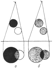
research, are characterized by strictly defined features, namely: number, position, shape, size, intensity, structure, nature of the contours, the presence or absence of mobility, dynamics over time.
X-ray methods of research
1. The concept of X-rays
X-rays are called electromagnetic waves with a length of approximately 80 to 10 ~ 5 nm. The longest-wavelength X-rays are covered by short-wavelength ultraviolet radiation, and the short-wavelength ones by long-wavelength Y-radiation. According to the method of excitation, X-ray radiation is divided into bremsstrahlung and characteristic.
The most common X-ray source is the X-ray tube, which is a two-electrode vacuum device. The heated cathode emits electrons. The anode, often called the anticathode, has an inclined surface in order to direct the resulting X-ray radiation at an angle to the axis of the tube. The anode is made of a highly heat-conducting material to remove the heat generated by the impact of electrons. The anode surface is made of refractory materials having a large atomic number in the periodic table, such as tungsten. In some cases, the anode is specially cooled with water or oil.
For diagnostic tubes, the pinpointness of the X-ray source is important, which can be achieved by focusing electrons in one place of the anticathode. Therefore, constructively, two opposite tasks have to be taken into account: on the one hand, electrons must fall on one place of the anode, on the other hand, in order to prevent overheating, it is desirable to distribute electrons over different parts of the anode. One of the interesting technical solutions is an X-ray tube with a rotating anode. As a result of deceleration of an electron (or other charged particle) electrostatic field atomic nucleus and atomic electrons of the substance of the anticathode, X-ray bremsstrahlung occurs. Its mechanism can be explained as follows. A moving electric charge is associated with a magnetic field, the induction of which depends on the speed of the electron. When braking, the magnetic induction decreases and, in accordance with Maxwell's theory, an electromagnetic wave appears.
When electrons decelerate, only part of the energy goes to create an X-ray photon, the other part is spent on heating the anode. Since the ratio between these parts is random, when a large number of electrons decelerate, a continuous spectrum of X-ray radiation is formed. In this regard, bremsstrahlung is also called continuous.
In each of the spectra, the shortest wavelength bremsstrahlung occurs when the energy acquired by an electron in the accelerating field is completely converted into the energy of a photon.
Short-wavelength X-rays usually have a greater penetrating power than long-wavelength ones and are called hard, while long-wavelength ones are called soft. Increasing the voltage on the x-ray tube, change the spectral composition of the radiation. If the cathode filament temperature is increased, then the electron emission and the current in the tube will increase. This will increase the number of X-ray photons emitted every second. Its spectral composition will not change. By increasing the voltage on the X-ray tube, one can notice the appearance of a line, which corresponds to the characteristic X-ray radiation, against the background of a continuous spectrum. It arises due to the fact that accelerated electrons penetrate deep into the atom and knock electrons out of the inner layers. Electrons from the upper levels pass to free places, as a result, photons of characteristic radiation are emitted. In contrast to optical spectra, the characteristic x-ray spectra of different atoms are of the same type. The uniformity of these spectra is due to the fact that the inner layers of different atoms are the same and differ only energetically, since the force effect from the nucleus increases with the increase in the ordinal number of the element. This circumstance leads to the fact that the characteristic spectra shift towards higher frequencies with increasing nuclear charge. This pattern is known as Moseley's law.
There is another difference between optical and x-ray spectra. The characteristic X-ray spectrum of an atom does not depend on the chemical compound in which this atom is included. So, for example, the X-ray spectrum of the oxygen atom is the same for O, O 2 and H 2 O, while the optical spectra of these compounds are significantly different. This feature of the X-ray spectrum of an atom served as the basis for the name characteristic.
characteristic Radiation always occurs when there is free space in the inner layers of an atom, regardless of the reason that caused it. So, for example, characteristic radiation accompanies one of the types of radioactive decay, which consists in the capture of an electron from the inner layer by the nucleus.
Registration and use of X-ray radiation, as well as its impact on biological objects, are determined by the primary processes of interaction of an X-ray photon with electrons of atoms and molecules of a substance.
Depending on the ratio of photon energy and ionization energy, three main processes take place
Coherent (classical) scattering. Scattering of long-wavelength X-rays occurs mainly without changing the wavelength, and it is called coherent. It occurs when the photon energy is less than the ionization energy. Since in this case the energy of the X-ray photon and the atom does not change, coherent scattering in itself does not cause biological action. However, when creating protection against X-ray radiation, one should take into account the possibility of changing the direction of the primary beam. This type of interaction is important for X-ray diffraction analysis.
Incoherent scattering (Compton effect). In 1922 A.Kh. Compton, observing the scattering of hard X-rays, discovered a decrease in the penetrating power of the scattered beam compared to the incident beam. This meant that the wavelength of the scattered X-rays was greater than that of the incident X-rays. The scattering of X-rays with a change in wavelength is called incoherent, and the phenomenon itself is called the Compton effect. It occurs if the energy of the X-ray photon is greater than the ionization energy. This phenomenon is due to the fact that when interacting with an atom, the energy of a photon is spent on the formation of a new scattered X-ray photon, on detaching an electron from an atom (ionization energy A) and imparting kinetic energy to an electron.
It is significant that in this phenomenon, along with secondary X-ray radiation (energy hv "of a photon), recoil electrons appear (kinetic energy £k of an electron). In this case, atoms or molecules become ions.
Photoelectric effect. In the photoelectric effect, X-ray radiation is absorbed by an atom, as a result of which an electron flies out, and the atom is ionized (photoionization). If the photon energy is insufficient for ionization, then the photoelectric effect can manifest itself in the excitation of atoms without the emission of electrons.
Let us list some of the processes observed under the action of X-rays on matter.
X-ray luminescence- the glow of a number of substances under X-ray irradiation. Such a glow of platinum-cyanogen barium allowed Roentgen to discover the rays. This phenomenon is used to create special luminous screens for the purpose of visual observation of x-rays, sometimes to enhance the action of x-rays on a photographic plate.
Known chemical action x-rays, such as the formation of hydrogen peroxide in water. A practically important example is the effect on a photographic plate, which makes it possible to detect such rays.
Ionizing action manifests itself in an increase in electrical conductivity under the influence of x-rays. This property is used in dosimetry to quantify the effect of this type of radiation.
One of the most important medical applications of X-rays is the transillumination of internal organs for diagnostic purposes (X-ray diagnostics).
X-ray method is a method of studying the structure and function of various organs and systems, based on a qualitative and / or quantitative analysis of an X-ray beam that has passed through the human body. The X-ray radiation that has arisen in the anode of the X-ray tube is directed to the patient, in whose body it is partially absorbed and scattered, and partially passes through. The image converter sensor captures the transmitted radiation, and the converter builds a visible light image that the doctor perceives.
A typical x-ray diagnostic system consists of an x-ray emitter (tube), an object of study (patient), an image converter and a radiologist.
For diagnostics, photons with an energy of about 60-120 keV are used. At this energy, the mass extinction coefficient is mainly determined by the photoelectric effect. Its value is inversely proportional to the third power of the photon energy (proportional to X 3), which manifests a large penetrating power of hard radiation and is proportional to the third power of the atomic number of the absorbing substance. The absorption of x-rays is almost independent of which compound the atom is in the substance, so one can easily compare the mass attenuation coefficients of bone, soft tissue, or water. A significant difference in the absorption of x-ray radiation by different tissues allows you to see images of the internal organs of the human body in a shadow projection.
A modern X-ray diagnostic unit is a complex technical device. It is saturated with elements of teleautomatics, electronics, electronic computers. A multi-stage protection system ensures radiation and electrical safety of personnel and patients.
